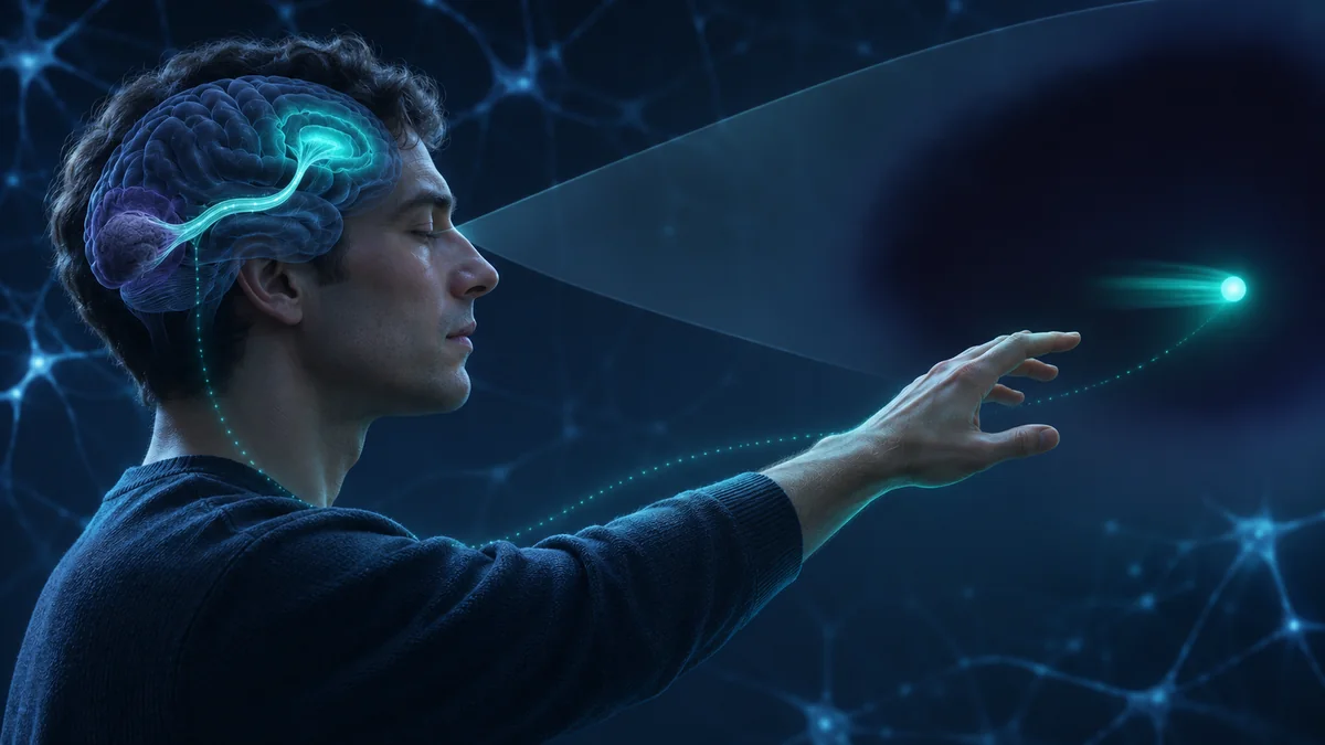
A patient says that nothing appeared in the left side of his vision. Asked to guess where a flash occurred, he points to it. Asked whether a line tilted left or right, he answers correctly more often than chance. He insists that he did not see it.
This is blindsight, a rare pattern found after damage to the primary visual cortex. It suggests that the brain can use visual information to guide decisions and actions even when a person reports no ordinary visual experience.
Seeing is not one process
Ordinary vision feels unified. A red ball appears, you know where it is, and you can reach for it. The brain separates these achievements across many systems. Some analyze colour, edges, motion, objects, space, and actions. Other processes make visual information available for report, confidence, and deliberate reasoning.
Blindsight exposes that separation. A person may lack the experience normally meant by “seeing” while retaining enough information to choose between locations or movements. The residual abilities do not amount to normal sight; whether some involve weak awareness remains disputed. Performance is limited, varies across patients and tasks, and often depends on high contrast or motion.
What damage causes blindsight?
Signals from the eyes travel through several routes. The dominant pathway passes through the lateral geniculate nucleus, or LGN, in the thalamus and then reaches the primary visual cortex, called V1, at the back of the brain. V1 sends organized information to other visual areas.
Damage to one side of V1 can create a blind region in the opposite side of visual space. This is often a hemianopia, a loss affecting half of the visual field. Damage to both sides can cause cortical blindness across most or all of the field even when the eyes still work.
V1 is central to conscious human vision, but it is not the only destination for visual signals. The superior colliculus helps orient the eyes and body. The pulvinar and LGN connect with extrastriate regions outside V1. These alternative routes can carry limited information about motion, location, contrast, or emotionally significant stimuli.
The experiment that named blindsight
In 1974, researchers studied a patient known as DB after surgery had removed part of his right visual cortex. Standard testing showed blindness in part of his left visual field. Yet when DB was asked to guess, he could localize flashes and distinguish some orientations and spatial patterns better than chance.
DB did not report seeing the stimuli in the ordinary way. Lawrence Weiskrantz and colleagues used the term blindsight to describe this mismatch between objective performance and acknowledged experience.
The method matters. If a researcher asks “Did you see something?”, the patient may say no. If the researcher forces a choice between two locations, chance performance would be 50 per cent. Repeated accuracy above that level shows that information influenced the answer even when the patient denied seeing it.
A single correct guess proves little. The evidence comes from many controlled trials, eye-position monitoring, comparisons with intact vision, and replication across tasks and patients.
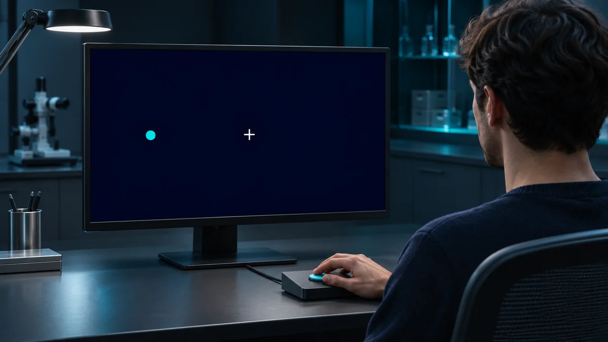
The participant describes whether there was any visual experience, including a faint or unusual sensation.
Across many trials, the researcher checks whether choices are more accurate than chance.
What can people do without ordinary sight?
Reported abilities include locating flashes, judging line orientation, discriminating motion direction, responding to colour or wavelength, and directing the eyes or hand toward a target. No patient keeps every ability, and the quality is usually far below normal vision.
Patient GY became one of the most studied cases. Experiments found above-chance motion and orientation discrimination in his blind field. Brain imaging showed that the same stimulus could produce different patterns depending on whether GY reported awareness. Under some conditions he described a vague feeling that something happened without normal visual qualities.
This led researchers to distinguish two broad forms. In type I blindsight, a person denies any awareness while performing above chance. In type II, the person reports a nonvisual feeling, movement sense, or awareness that something occurred without clearly seeing it.
Another patient, known as TN, had extensive bilateral damage to V1. In a striking 2008 demonstration, he navigated around obstacles in a corridor without a cane or assistance while reporting no visual awareness of the obstacles. The result showed useful visually guided behaviour, but it did not establish that TN formed a conscious picture of the corridor.
Can an unseen face affect emotion?
Some patients can discriminate emotional facial expressions presented in a blind field better than chance. Their facial muscles, skin conductance, or amygdala activity can also differ for emotional expressions they say they did not see. This pattern is called affective blindsight.
The proposed route emphasizes fast subcortical pathways involving the superior colliculus, pulvinar, and amygdala. These systems may prioritize biologically relevant signals before detailed conscious recognition.
Affective blindsight does not mean the brain reads every hidden emotion accurately. Effects depend on stimulus properties and experimental design. Low spatial frequencies, which carry broad facial structure rather than fine detail, appear especially important in some studies.
How can vision bypass V1?
There is probably no single blindsight pathway. Different spared routes may support different abilities.
- LGN to hMT+: A pathway from the lateral geniculate nucleus to the motion-sensitive area hMT+ can bypass V1 and support motion discrimination.
- Superior colliculus routes: These can support rapid orienting, eye movements, attention, and some visually guided action.
- Pulvinar connections: These may contribute to attention and emotional processing through links with higher visual and temporal areas.
- Spared tissue: Small surviving parts of V1 can contribute in some patients, though they do not explain every well-controlled case.
In 2015, Sara Ajina and colleagues compared people with V1 damage who did or did not show blindsight. An intact structural connection between the LGN and hMT+ was associated with residual visual ability. Later work found that this functional connection, rather than a pulvinar-to-hMT+ route, was especially important for motion blindsight.
A 2025 systematic review concluded that human and monkey evidence supports several parallel pathways. Which route matters depends on lesion extent, individual anatomy, the stimulus, and time since injury.
Dashed border: V1 injury disrupts this route.
Is blindsight really unconscious?
This is the central dispute. The traditional interpretation says that visual information guides behaviour without visual consciousness. A competing view says the experience may be extremely weak, degraded, or unusual rather than absent.
Subjective reports require a decision threshold. A patient may only say “seen” when experience is clear. A faint signal below that threshold can still improve forced-choice accuracy. If so, blindsight could partly reflect conservative reporting rather than pure unconscious perception.
Researchers have tried to improve the measurement. Confidence ratings ask how certain a person is. Post-decision wagering asks participants to risk more when they believe an answer is correct. In a 2007 study, participants with blindsight and other implicit-learning effects sometimes failed to bet optimally despite accurate choices, supporting a gap between performance and knowledge of performance.
Yet wagering is not a direct consciousness meter. People may avoid risk, misunderstand probabilities, or separate confidence from visual experience. Studies of GY found that confidence could carry information even when he denied visual awareness. These results show why one yes-or-no report cannot settle the question.
The strongest position is measured: blindsight demonstrates a major dissociation between visual processing and ordinary reportable vision. Some cases appear consistent with no awareness; others include weak or nonvisual experience. The category may contain several phenomena.
A visual scene with identifiable features.
BehaviourCan usually identify and act on what is seen.
No acknowledged awareness of the stimulus.
BehaviourAbove-chance performance on a tested visual task.
A vague sense that something occurred, without ordinary seeing.
BehaviourResidual ability on a tested visual task.
Attention and action without awareness
Blindsight also separates consciousness from attention. Spatial cues can speed discrimination in the blind field even when the stimulus is not reported. Attention can enhance processing without being sufficient for awareness.
Action may use visual information differently from conscious description. Reaching, orienting, or navigating can succeed when verbal identification fails. Patient TN could sometimes localize a target when responding with a goal-directed movement but not with an arbitrary button response.
This does not prove two completely independent visual brains. The systems interact. It shows that conscious recognition is only one use of visual information.
What blindsight teaches about consciousness
First, accurate behaviour is not sufficient evidence of conscious experience. A brain can transform sensory input into a useful response without producing ordinary reportable vision.
Second, verbal report is not a perfect measure either. Faint experiences, unusual sensations, and decision criteria complicate any clean division between conscious and unconscious.
Third, V1 appears crucial for the rich, stable visual world humans normally experience, while other routes can preserve fragments of function. Conscious vision depends on how information is integrated and made available across networks, not on the mere presence of a visual signal.
Finally, blindsight cautions against equating intelligence or performance with sentience. This matters when discussing animals, brain injury, and artificial systems. Successful discrimination tells us what a system can use. It does not by itself tell us what, if anything, the system experiences.
Clinical limits and rehabilitation
Blindsight is a research description, not a diagnosis that restores normal vision. Most people with visual-field loss cannot rely on residual processing in daily life, and laboratory performance may require hundreds of trials.
Training can improve detection or discrimination in some blind-field locations. Researchers are testing whether preserved pathways predict recovery and whether repeated motion training expands useful function. Gains can be specific to the trained location and task.
Any assessment or rehabilitation belongs with vision and neurological specialists. The scientific value of blindsight does not guarantee a practical ability for an individual patient.
How researchers test awareness carefully
A rigorous blindsight experiment first maps the damaged visual field and confirms that the stimulus remains inside it. Eye tracking matters because a small eye movement could place the target in a sighted region. Researchers then vary contrast, duration, location, and motion while comparing forced choices with awareness reports.
Signal detection methods separate sensitivity from response bias. A participant who almost always says “nothing” may still distinguish targets, but the analysis must show that performance cannot be explained by a preference for one answer. Trials with no stimulus help reveal guessing strategies and false alarms.
Lesion mapping is equally important. Residual islands of V1, incomplete damage, and recovery can imitate or contribute to blindsight. Modern studies combine structural imaging, diffusion imaging of white-matter pathways, functional brain activity, eye position, and detailed psychophysics.
Even this does not directly observe experience. Science compares reports, confidence, choices, physiology, and neural activity. When those measures separate, researchers gain evidence about the components of consciousness, but interpretation remains tied to the limits of each measure.
Common questions
Are people with blindsight completely blind?
Usually they have a blind region caused by cortical damage. Some have bilateral cortical blindness. Residual abilities are limited and do not equal normal conscious sight.
Do their eyes still work?
Often the eyes and early pathways still receive light. The major damage is in V1 or its inputs, which prevents normal cortical vision in part of the field.
Can someone with blindsight drive?
Laboratory discrimination does not establish safe driving ability. Visual-field requirements and clinical assessments determine fitness to drive.
Is blindsight proof of unconscious perception?
It is strong evidence that visual information can guide behaviour without ordinary reported vision. Researchers still debate whether some cases involve faint or unusual conscious experience.
Can healthy people have blindsight?
The clinical phenomenon follows brain damage. Experiments using masking or temporary disruption can produce related dissociations in healthy participants, but they are not identical to neurological blindsight.
Sources and further reading
Sanders and colleagues, 1974, The Lancet. Original localization and discrimination findings in patient DB.
Sahraie and colleagues, 1997, PNAS. Brain activity associated with conscious and unreported visual processing in GY.
Sahraie, Weiskrantz, and Barbur, 1998, Behavioural Brain Research. Awareness and confidence during motion discrimination.
Persaud, McLeod, and Cowey, 2007, Nature Neuroscience. Post-decision wagering as a measure of awareness.
de Gelder and colleagues, 2008, Current Biology. Obstacle navigation after bilateral loss of V1.
Tamietto and de Gelder, 2008, Neuropsychologia. Emotional processing without reported visual experience.
Ajina and colleagues, 2015, eLife. Human blindsight and the LGN-to-hMT+ pathway.
Derrien and colleagues, 2022, Neuroscience of Consciousness. Review of blindsight types and implications for consciousness theories.
de Gelder, 2025, Cerebral Cortex. New naturalistic observations of residual awareness in cortical blindness.
Balali and colleagues, 2025, Basic and Clinical Neuroscience. Systematic review of pathways mediating blindsight.